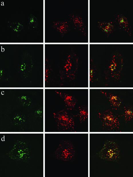
Fig. 2.
Internalized LFn-GFP does not completely localize with the endocytic or secretory pathways. HeLa cells incubated for 1 hr with LFn-GFP in the absence of PA were stained with markers for early endosomes (EEA-1; a), late endosomes (Lamp-1; b), lysosomes (Lamp-2; c), and the Golgi apparatus (Ab-1; d) and visualized by confocal microscopy. (Right) Overlay of red and green staining. (Center) Red organelle antibody staining. (Left) Green fluorescence of LFn-GFP.

