Fig. 1.
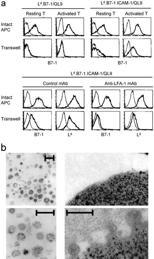
TCR-mediated absorption of membrane vesicles. (a) Absorption of B7-1 by purified naïve or activated CD8+ 2C cells after incubation for 1 h with Ld.B7-1 or Ld.B7-1.ICAM-1 Dros APCs loaded with QL9 peptide at 10 μM; activated T cells were prepared by preculturing cells for 12 h with PMA plus ionomycin. T cells and APCs were either cultured together (intact APCs) or separated from each other by placing APCs in a transwell (pore size 3 μm); Dros cells are large (>20 μm) and were unable to pass through the Transwell membrane. After culture, T cells were stained for B7-1, Ld, and CD8 and then examined by flow cytometry. The data show staining of gated CD8+ cells. (a Upper) B7-1 staining of resting vs. activated 2C cells after culture with Ld.B7-1 vs. Ld.B7-1.ICAM-1 Dros APCs. (a Lower) B7-1 and Ld staining of activated 2C cells cultured with Ld.B7-1.ICAM-1 Dros APCs in the absence or presence of anti-LFA-1 mAb (5 ng/ml). (b) Morphology of membrane vesicles. Culture supernatants from Ld.B7-1.ICAM-1 Dros APCs were depleted of cell debris, then ultracentrifuged. (Left) Electron microscopic view of pelleted material is shown at low (Upper) and high (Lower) magnification. (Bar, 200 nm.) Much of the material in the pellet shows the morphology of membrane vesicles; cell debris, more prominent in other fields, is also present. (Right) Electron microscopic view of magnetic beads that were coated with anti-ICAM-1 mAb (Lower) or an Ig isotype-matched control mAb (Upper) before incubation with the above membrane vesicles.

