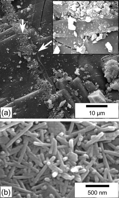
Fig. 1.
SEM images (secondary electrons) of the surface of the Tataouine meteorite. (a) At the center of the image, a filament (the arrow and Inset show the filament at a higher magnification) is bordered all along its axis with clusters of nanobacteria-like rods. The euhedral crystal at the bottom right of the picture is a chromite. (b) Close-up view of the area denoted by a rectangle in a. The nanobacteria-like rods are 80 nm wide and a few hundreds of nanometers long. They display round edges.

