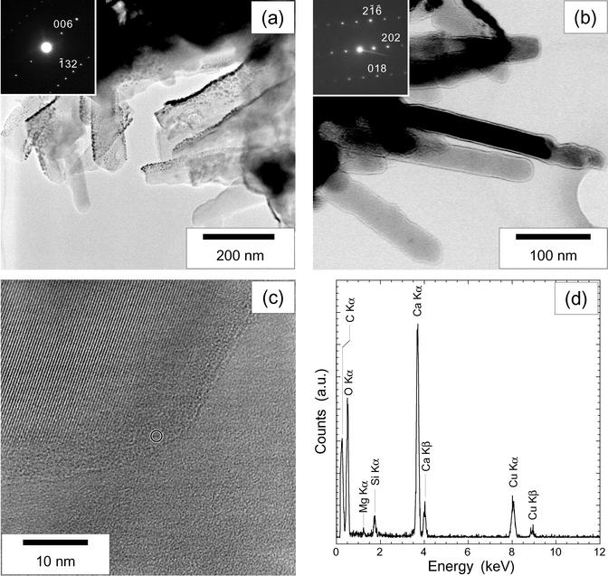
Fig. 3.
TEM characterization of the clusters of nanobacteria-like rod-shaped forms from the surface of the Tataouine meteorite. (a) Image of a cluster. (Inset) Electron diffraction pattern obtained on a single rod (zone axis [310]). Tiny dense spots in the image correspond to the gold-coating particles. (b) Image of another cluster. At this magnification, the edges of nanobacteria-like rods seem to be smooth. A strong contrast at the borders of the nanobacteria-like rods reinforced by overfocusing can be noticed. Zone axis [18-1] is shown. (c) High-resolution image of the border of a nanobacteria-like rod. The crystallized core showing lattice fringes is facetted and surrounded by an amorphous layer. The circle indicates the size of the EDX probe that was used. (d) EDX analysis obtained from the rod showing the background signal of the silicon-coated copper grid on which the samples have been deposited. Calcium, carbon, and oxygen are measured in both the core and the amorphous layer of the nanoforms. No other element has been evidenced. a.u., arbitrary units.

