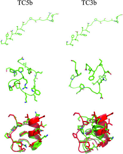
Fig. 1.
Sample structures from the TC5b (Left) and TC3b (Right) simulations. Simulated structures are shown in green. (Top) Initial extended conformation of each peptide. (Middle) Peptide structures after 50 ps of 298-K molecular dynamics (MD) equilibration. (Bottom) Final 298-K conformation for each peptide after the 4-ns replica-exchange run. The simulated structures are shown superimposed on the first conformation of the TC5b NMR ensemble (shown in red). The structures were superimposed based on Cα RMSD. These images were generated with the pymol program.

