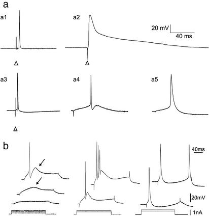
Fig. 2.
Emerging intrinsic membrane properties of eGFP-expressing myotubes. (a) Action potential characteristics of 5 types of muscle cell. (a1) Tibialis skeletal muscle cell; (a2) ventricular myocyte; (a3) eGFP-expressing myotube grafted for 28 days in the tibialis muscle; (a4) eGFP-expressing myotube grafted for 28 days in infarcted myocardium; (a5) eGFP-expressing myotube maintained in culture for 28 days. Whether grafted or in culture, eGFP-expressing myotubes fired brief action potentials (a3, a4, a5) in comparison with myocytes (a2). Note that the action potential of the myotubes grafted in infarcted myocardium (a4) is followed by a fast afterhyperpolarization. The action potentials were evoked either with extracellular stimulations (arrow heads point to the stimulation artifact) or with intracellular depolarizing current injections. (b) Firing properties of eGFP-expressing myotubes recorded in infarcted myocardium (Left and Center) or in culture (Right). (Left) Subthreshold depolarizing current pulses evoked a slow voltage-dependent depolarizing hump (arrow, middle trace) on top of which an action potential was triggered on higher stimulation (top trace). (Center) In another cell, a burst of action potentials was emitted on the slow depolarizing hump and followed by a slow afterhyperpolarization. (Right) Myotubes in culture do not fire bursts of action potentials.

