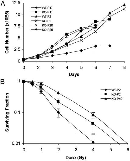
Fig. 2.
Growth of MEF cells. MEF cultures were established from both PKCI+/+ (WT) and PKCI-/- (KO) mice by using 13.5-day embryos. The cells were serially passaged in DMEM with 10% FBS. (A) Growth curves were carried out with WT and KO MEF cells at passage (P) 2, 10, 20, and 25. (B) Relative sensitivity of WT and KO MEF cells to ionizing radiation. Exponentially growing early-passage 2 (P2) WT and KO MEF cells and late-passage (P42) immortalized KO MEF cells were treated with ionizing radiation ranging from 0.5 to 6 Gy. The numbers of colonies were then determined and plotted as the surviving fraction (SF). SF = PE of irradiated cells/PE of control unirradiated cells, where plating efficiency (PE) = number of colonies/number of cells seeded.

