FIG. 6.
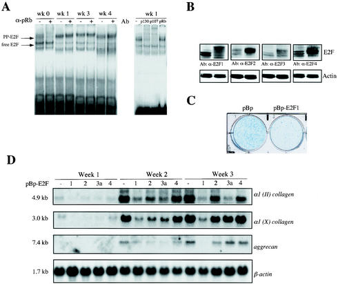
Ectopic expression of E2F transcription factors alters differentiation of chrondogenic ATDC5 cells. (A) Electrophoretic mobility shift analysis on extracts from prechondrogenic cells (week [wk] 0) and early (week 1) and late differentiating chondrocytes (week 3 and 4). The positions of the free E2F-DNA and pocket protein (PP)-E2F-DNA complexes are indicated by the arrows to the left of the gel. Supershift analyses with specific antibodies (Ab) against pRb (α-pRb) (21C9), p107 (C-18), and p130 (C-20) indicate the presence of distinct pocket protein complexes. (B) Western blot analysis shows expression of different E2F species in mock-infected pBabepuro ATDC5 cells (left lane of each blot) or polyclonal populations of ATDC5 cells infected with pBp-HA-E2F1, pBp-HA-E2F2, pBp-HA-E2F3a, or pBp-HA-E2F4 (right lane of each blot). Antibodies (Ab) against E2F1 (α-E2F1), E2F2, E2F3a, and E2F4 were used. (C) Cultures of polyclonal cells infected with pBabepuro (pBp) and pBp-HA-E2F1 were stained with Alcian blue after differentiation for 15 days, which detects differentiated cartilage nodules. (D) Expression of cartilage markers at different phases of chondrocyte differentiation in ATDC5 cells, which stably overexpress pBabepuro (pBp), pBp-HA-E2F1, pBp-HA-E2F2, pBp-HA-E2F3a, and pBp-HA-E2F4 (−, 1, 2, 3a, and 4, respectively). Total RNA was isolated at the indicated times and subjected to Northern blot analysis. Filters were serially hybridized with cDNA probes for type II collagen [α1 (II) collagen]), type X collagen [α1 (X) collagen], and aggrecan genes and β-actin, which serves as a loading control. Transcript sizes are indicated to the left of the blots.

