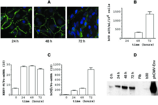
FIG. 2.
Expression of HERV-W env mRNA and glycoprotein is colinear with primary cytotrophoblast differentiation. (A) Desmoplakin immunodetection after 24, 48, and 72 h of culture of trophoblast cells isolated from a term placenta. Positive immunofluorescent staining is observed only in aggregated cytotrophoblasts (24 h) or in cytotrophoblasts in contact with the syncytiotrophoblast (48 h); staining has disappeared in the fused syncytiotrophoblast characterized by multiple nuclei (72 h). Nuclei were labeled with DAPI (blue fluorescence). (B) Levels of hCG (expressed in milli-international units per milliliter per 106 cellules) secreted into the culture medium at the indicated times and characterizing the observed differentiation. Since cells were plated in triplicate (see Materials and Methods), hCG levels were determined for each plate. Results are expressed as the mean and SEM of the three culture dishes. (C) Env-W and hCG-mRNA expression in cytotrophoblasts (CT0) and during their in vitro differentiation for 3 days. mRNA data are expressed as the level of each marker mRNA normalized by RPLPo mRNA levels (Po). Two culture dishes were pooled for each determination, and transcripts were assayed in duplicate. (D) Western blot analysis of Env glycoprotein detected by the anti-SU polyclonal antibody during trophoblast differentiation (0 to 72 h). Fibroblast protein extract was used as a negative control (Fib), and a BeWo cell line (B30) and env-transfected TE 671 cells (phCMV-Env) were used as positive controls. The whole figure (A through D) illustrates an experiment which is representative of four.

