FIG. 3.
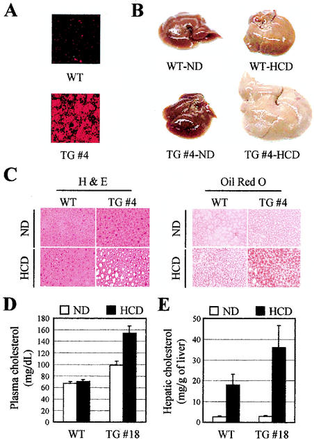
Morphology and histology of livers from DN2-TG (TG) and wild-type (WT) mice on high-cholesterol diets. (A) Expression of DN2 was assessed with indirect immunofluorescence with αHA antibody. TG #4 indicates the DN2-TG line no. 4 (Table 1). (B) Gross morphology of livers from male DN2-TG and wild-type mice fed chow supplemented with 2% cholesterol (HCD, high-cholesterol diet) for 90 days. ND, normal chow diet. The development of fatty livers in the DN2-TG mice is evident after 7 days on the high-cholesterol diet. (C) Sections from livers shown in panel B were prepared for histology and stained with oil red O, as indicated. The unstained vacuoles visible in the hematoxylin and eosin (H & E)-stained sections of the livers from DN2-TG mice on the high-cholesterol diet stain positive (red) for lipids with oil red O. (D and E) Measurements of plasma and hepatic cholesterol quantitated enzymatically from extracts of the livers shown in panel B. All values are expressed as means plus standard errors of the mean; n = 5.

