FIG. 8.
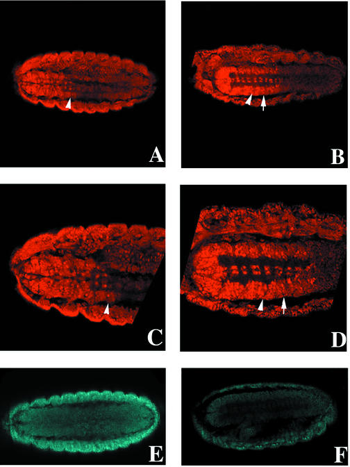
EXD expression in the CNSs of Drosophila wild-type and zipper mutant embryos. EXD was visualized in wild-type (A and C) and zipper mutant (B and D) embryos with an anti-EXD antibody. The arrowheads indicate the boundary between the third thoracic (T3) and first abdominal (A1) segments. Anterior is to the left. (A) Ventral view of EXD staining in the CNS of a stage 15 wild-type embryo. The boundary between cells expressing high nuclear versus low cytoplasmic EXD is located at the T3-A1 border. (C) Closer examination of the thoracic CNS shows that most cells express EXD at intermediate to high levels in both the cytoplasm and nucleus, or mainly in the nucleus. In the abdominal CNS, most cells contain low levels of strictly cytoplasmic EXD. (B) EXD staining in the CNS of a stage 15 zipper mutant embryo. The boundary between nuclear versus cytoplasmic EXD (arrow) is shifted posteriorly to encompass A1. (D) Closer view of CNS cells in a zipper mutant. Cells in A1 (between the arrowhead and the arrow) express high levels of EXD in both the cytoplasm and nucleus. Several “z” planes were examined for each embryo to ensure that differences in EXD expression were not artifacts of the plane of section (data not shown). Four out of four zipper mutant embryos for which ventral views of the CNS were possible displayed the same posteriorized EXD nuclear localization. (E and F) The expression level of ZIPPER in a zipper mutant embryo (F) is greatly reduced compared to that in a wild-type embryo (E).

