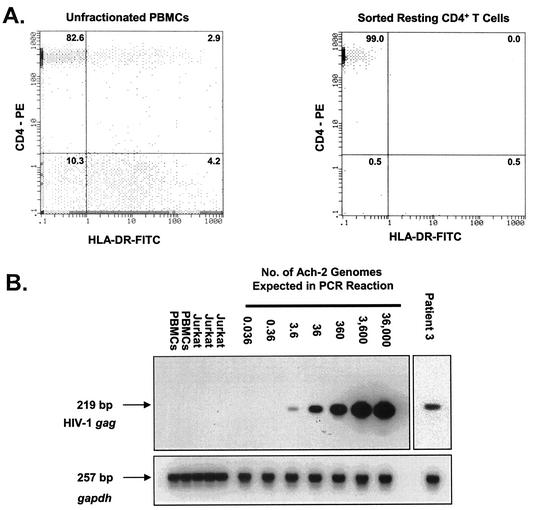
FIG. 1.
Quantitative measurements of HIV-1 proviral DNA present in highly purified resting CD4+ T lymphocytes. (A) Isolation of resting CD4+ T lymphocytes from patients on HAART. The results of flow cytometric analysis of CD4 and HLA-DR expression on a gated lymphocyte population in unfractionated PBMCs (left panel) and on an ungated sorted population of highly purified resting CD4+ T lymphocytes (right panel) are shown. The numbers are the percentages of cells expressing CD4 and/or HLA-DR. (B) Quantification of HIV-1 DNA in highly purified resting CD4+ T lymphocytes from patients on HAART. To generate a standard curve, ACH-2 cells carrying a single integrated copy of the HIV-1 genome were diluted with HIV-1-negative PBMCs. Isolated DNA was amplified for HIV-1 gag as described in Materials and Methods. To control for DNA quality, the cellular gapdh gene was amplified in parallel. PCR products were confirmed by Southern hybridization using gene-specific probes.

