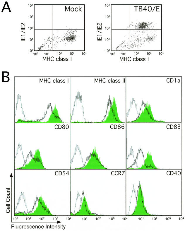
FIG. 4.
Human CMV infection alters the cell surface expression of key molecules involved in mDC function. (A) Dot plot showing expression of MHC class I molecules on the surface of mock infected (left) or CMV strain TB40/E-infected LC-derived mDCs (right) at day 2 p.i. Cells were stained with a PE-conjugated anti-MHC class I monoclonal antibody (x axis), as well as with an FITC-conjugated anti-IE1/IE2 monoclonal antibody (y axis). (B) Flow cytometry analysis of expression levels of MHC class I, MHC class II, CD1a, CD80, CD86, CD83, CD54, CCR7, and CD40 on the surface of mock-infected and CMV strain TB40/E-infected LC-derived mDCs at day 2 p.i. Filled histograms, mock-infected cells; open histograms, TB40/E-infected cells; gray line, isotype controls.

