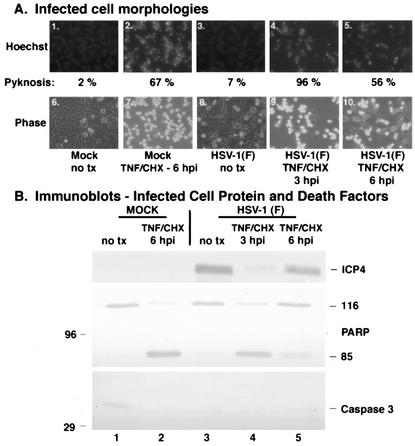
FIG. 1.
Infected cell proteins produced by wild-type HSV-1(F) infection by 6 hpi block TNF-α-induced apoptosis. Fluorescence and phase-contrast images of infected cells (A) and immune reactivities of infected cell proteins and cellular death factors (B) were obtained as described in Materials and Methods. HEp-2 cells were infected with HSV-1(F) (MOI = 5) or mock infected, TNF-α plus CHX was added at 3 or 6 hpi, Hoechst dye was added 60 min prior to harvesting, and the number of cells (percent) with pyknotic nuclei (Pyknosis) was determined from two independent experiments. Infected whole-cell extracts were prepared at 18 hpi (TNF-α-plus-CHX treatments) or 24 hpi (without treatment), separated in a denaturing gel, and transferred to nitrocellulose, and immunoblotting was performed with anti-ICP4 and anti-PARP antibodies. 116 and 85 refer to full-length and processed PARP, respectively. “no tx” refers to no TNF-α-plus-CHX treatment. Magnification, ×40.

