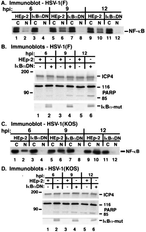
FIG. 10.
Apoptosis in IκBαDN cells following HSV-1(F) (A and B) and HSV-1(KOS1.1) (C and D) infection. Duplicate sets of HEp-2 or IκBαDN cells were infected with HSV-1(F) or HSV-1(KOS1.1) (MOI = 10) and at 6, 9, and 12 hpi, cytoplasmic (C) and nuclear (N) and whole-cell extracts were prepared, separated in denaturing gels, transferred to nitrocellulose, and probed with anti-NF-κB, anti-ICP4, anti-PARP, and anti-IκBα antibodies as described in Materials and Methods. IκBα-mut denotes the mutant protein, as the native form gets degraded and is not visible on the immunoblot. Anti-ICP4 and anti-IκBα immune reactivities were detected using alkaline phosphatase methods, while anti-PARP and anti-NF-κB reactivities were detected using a chemiluminescence technique.

