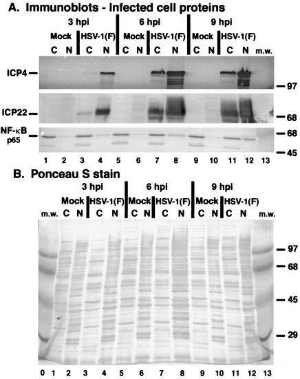
FIG. 2.
Immune reactivities (A) and Ponceau S staining (B) of infected cell extracts indicate that NF-κB partitions in the nuclear fraction of HSV-1(F)-infected cells. HEp-2 cells were mock infected or infected with HSV-1(F) (MOI = 5), cytoplasmic (C) and nuclear (N) extracts were prepared at 3, 6, and 9 hpi, and infected cell proteins were separated in a denaturing gel and transferred to nitrocellulose as described in Materials and Methods. Immune reactivities with anti-ICP4 and anti-NF-κB antibodies were determined using alkaline phosphatase detection methods, while reactivities with anti-ICP22 antibody were determined by using a chemiluminescence technique. The anti-NF-κB recognizes the p65 subunit of the protein. Ponceau S staining prior to immunoblotting was used to demonstrate equal protein loadings and to show that efficient separation of nuclear and cytoplasmic fractions had occurred. The sizes of molecular mass (m.w.) markers (in kilodaltons) are shown in the righthand margins.

