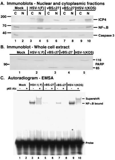
FIG. 7.
NF-κB does not translocate to the nuclei of apoptotic HSV-1(vBSΔ27)-infected cells and does not bind DNA. Immune reactivities of nuclear and cytoplasmic fractions (A) and whole-cell extract (B) of infected cell proteins and autoradiographic images (C) of radiolabeled DNA-protein complexes are shown. HEp-2 cells were mock infected or infected with HSV-1(F), HSV-1(vBSΔ27), or HSV-1(KOS1.1) (MOI = 10) and at 12 hpi, cytoplasmic (C) and nuclear (N) extracts were prepared, separated in denaturing gels, transferred to nitrocellulose, and probed with anti-ICP4, anti-caspase 3, and anti-NF-κB antibodies as described in Materials and Methods. To ensure consistency in this study, two separate infections were performed with two independent stocks of HSV-1(vBSΔ27) (lane 5 to 8). All immune reactivities were detected using an alkaline phosphatase technique. The locations of molecular mass markers (in kilodaltons) are shown in the left margin. Duplicate sets of HEp-2 cells were mock infected or infected with HSV-1(F), HSV-1(KOS), and HSV-1(vBSΔ27) (MOI = 10), and at 12 hpi, nuclear or whole-cell extracts were prepared. Whole-cell extracts were used for immunoblotting and probed with an anti-PARP antibody. Approximately 3 μg of nuclear extract was reacted with a 32P-labeled NF-κB site probe and electrophoresed in a nondenaturing polyacrylamide gel, and the gel was dried and exposed to autoradiographic film as described in Materials and Methods. A control anti-NF-κB p65 antibody was added (+) prior to addition of the labeled DNA probe. NF-κB that bound to the DNA probe and that supershifted by the anti-p65 antibody is indicated.

