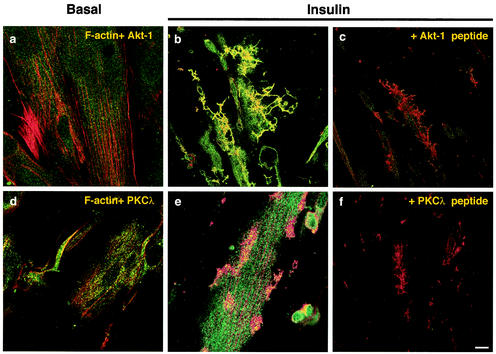
FIG. 2.
The PI3-K effector, Akt-1, but not aPKC, colocalizes with insulin-induced actin structures. Serum-deprived (4 h) L6 myotubes were untreated (a and d) or stimulated with 100 nM insulin for 10 min at 37°C (b, c, e, and f), followed by fixation and permeabilization. F-actin was labeled with rhodamine-conjugated phalloidin. Akt-1 (a to c) or PKCλ (d to f) proteins were stained with specific polyclonal antibodies, followed by Alexa 488-conjugated secondary antibodies, as described in Materials and Methods. Competitive inhibition of Akt-1 (c) and PKCλ (f) using a 10-fold molar excess of blocking peptides is also shown. Yellow regions indicate regions of remodeled actin filaments with colocalized Akt-1 or PKCλ protein. Shown are images of the dorsal plane of basal cells and dorsal extension planes of insulin-stimulated cells, as defined in the legend for panel a of Fig. 1A. The images are representative of four experiments. Bar, 10 μm.

