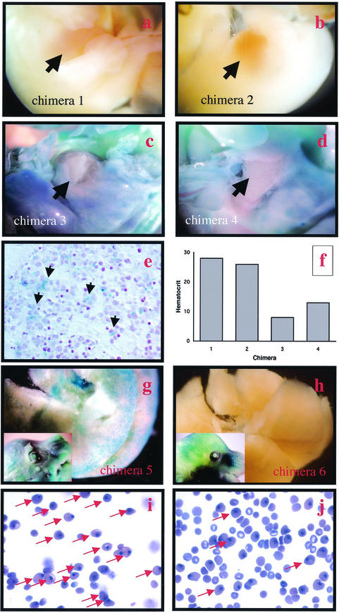
FIG.4.
Chimeric embryos with livers with large contributions of ES cells are anemic. (a to d) Four E14.5 nrf1lacZ/lacZ chimeric littermates whole mount stained with X-Gal. (e) Cross section of an E14.5 chimeric fetal liver stained with X-Gal showing intense blue staining of hepatocytes. (f) Hematocrits of the chimeric mice shown in panels a to d. Note that chimeras 3 and 4 (c and d), which showed blue staining in their livers (arrows), have low hematocrits. (g and h) E16.5 nrf1lacZ/lacZ chimeric littermates whole mount stained with X-Gal. (i and j) Blood smears of E16.5 nrf1lacZ/lacZ chimeric littermates. (i) Note that the blood of chimera 5 (g), which showed intense blue staining in the liver, contains large number of yolk sac-derived nucleated red cells (arrows) and few nonnucleated red cells of fetal liver origin. (j) Blood of chimera 6 (h), which showed intense X-Gal staining in the embryo (inset) but not the liver, contains both nucleated erythrocytes and nonnucleated red cells.

