FIG.6.
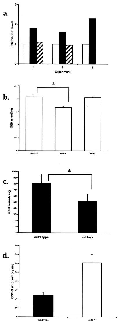
Nrf1-deficient fetal livers exhibit increased oxidative stress. (a) ROS levels of E13.5 liver samples from wild-type (white bars), nrf1−/− (black bars), and nrf2−/− (hatched bars) mice as determined by flow cytometric analysis of intracellular DCF fluorescence. Results from three experiments are shown. Levels are expressed relative to the signal for the wild-type sample in each experiment, which was arbitrarily assigned a value of one. In experiment 3, primary hepatocytes were used; fetal liver cells were cultured overnight on collagen-coated plates to remove nonadhering hematopoietic cells. (b) GSH levels in the livers of wild-type and nrf1+/− (control) (n = 8), nrf1−/− (n = 4), and nrf2−/− (n = 4) mice. (c) GSH levels of primary hepatocytes from E13.5 control (n = 5) and nrf1−/− (n = 3) fetuses. (d) GSSG levels of wild-type and nrf1−/− livers. Mean values ± standard deviations (error bars) are shown. Values that were statistically significantly different (P < 0.05 by Student's t test) are indicated by an asterisk.

