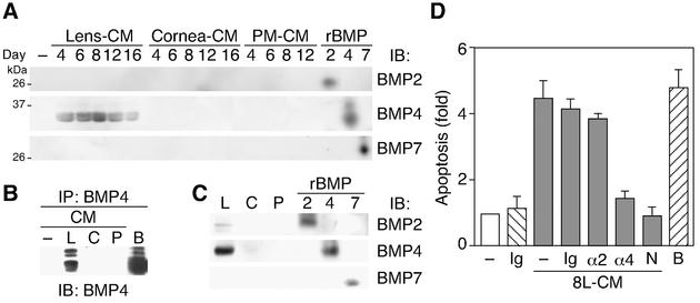
FIG. 6.
Identification of BMP4 as a factor required for Lens-CM-induced apoptosis of HUVE cells. (A and B) Developmental enhancement of BMP4 secretion from lens. Organ-CM (A) or protein immunoprecipitated by the anti-BMP4 antibody from organ-CM of day 8 rats (B) was stained with the specific antibodies. (C) Expression profile of BMP4 in organs. Lens, cornea and PMs were lysed and subjected to immunoblotting analysis using the specific antibodies. Results (A to C) are representative ones of at least three independent experiments. Anti-BMP4 antibody used for immunoblotting (A and C) and immunoprecipitation (B) was obtained from R&D Systems. In (B), anti-BMP4 antibody from Santa Cruz was used for immunoblotting. (D) Inhibitory effect of neutralizing antibody to BMP4 on Lens-CM-induced apoptosis. HUVE cells were cultured in M199 containing 5% FBS with or without Lens-CM from day 8 rats (8L-CM). Before use, the 8L-CM was immunodepleted with or without (−) the normal mouse IgG (Ig), the anti-BMP2 antibody (α2) and the anti-BMP4 antibody (α4). After 24 h of culture, TUNEL-positive cells were counted. HUVE cells cultured with Lens-CM and Noggin (250 ng/ml, N) were used as a positive control. Relative apoptotic cell numbers are shown as the mean ± standard error from several independent experiments. Abbreviations: B, BMP4; C, cornea; L, lens; N, Noggin; P, PM.

