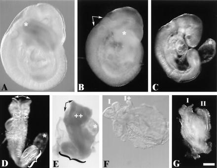
FIG. 5.
Phenotype of H1cH1dH1e−−−/−−− triple-homozygous mutant embryos recovered on E9.5. Approximately 60% of the expected number of homozygous mutant embryos are recovered at E9.5. The broad spectrum of abnormalities observed in recovered homozygous mutant embryos fall into three general classes. In the most predominant category (∼50%), representatives of which are shown in panels B and C, are embryos that were slightly developmentally delayed relative to their littermates (a representative wild-type embryo is shown in panel A) and presented a variety of incompletely penetrant abnormalities. Abnormalities observed in this class of mutants included occlusion of the rhombencephalic ventricle (arrows in panel B), a short tail (compare asterisks at the tail tips in panels A and B), and failure of the allantois to fuse with the chorion (asterisk in panel C). Mutant embryos falling into a second class, representatives of which are shown in panels D and E, are very developmentally delayed compared to their littermates and again variably presented developmental perturbations, including splayed anterior neural tubes (arrows in panels D and E), regions of excess tissue (++ in panel E), pericardial expansion (asterisk in panel E), failure of chorioallantoic fusion (asterisk in panel D), and caudal dysgenesis (braces in panels D and E). Mutant embryos in a third class are severely abnormal (F and G). They had not progressed past an early somite stage and in each case showed morphological evidence of partial (I and I+ in panel F) or complete (I and II in panel G) axis duplication. Embryos are viewed from the right side in panels A to C and E, from nearly dorsal in panel D, from ventral in panel F, and from a midline between the axes in panel G. Scale bar = 300 μm.

