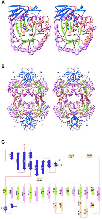
FIG. 4.
Structure of HYDBp. (A) Ribbon diagram (10) showing the overall structure of the HYDBp monomer viewed from the catalytic active site. (B) Tetramer of HYDBp showing the intersubunit interfaces. (C) Secondary structural topology of HYDBp. α-Helices are shown as cylinders; β-strands are shown as arrows. The amino acid residue ranges of the secondary structural elements are illustrated. The β-strands of the small β-sheet domain are shown in blue; the β-strands of the TIM barrel are shown in green; α-helices of the TIM barrel are shown in lavender; and the other α-helices are shown in pink. The two zinc ions at the catalytic site are illustrated as yellow spheres.

