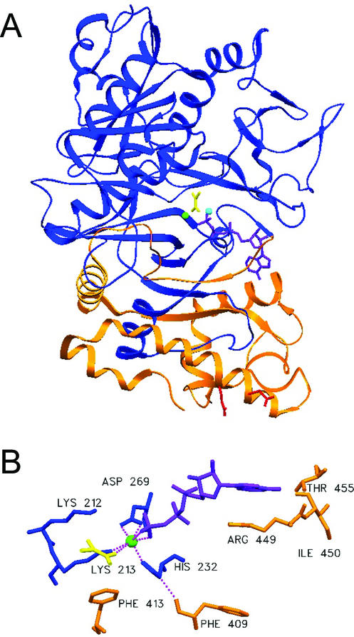
FIG. 5.
(A) PCK molecule showing C-terminal domain cleaved by trypsin (orange). The rest of PCK is colored blue. ATP and pyruvate are purple, Ca2+ is green, Mg2+ is cyan, and residues Glu508 and Glu511 are red. (B) View of residues interacting with Ca2+ in blue and C-terminal Phe413 and Phe409 (both orange) interacting with Lys213 and with His232, respectively.

