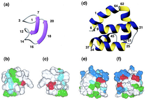
FIG. 1.
Solution structures of the circular bacterial proteins for which three-dimensional structures have been determined. (a) Ribbon representation of MccJ25 (PDB code 1HG6), with the β-strands shown as arrows. (b and c) Surface diagrams of MccJ25, with b in the same orientation as a and c rotated 180° about the y axis. White, green, and red represent hydrophobic, hydrophilic, and negatively charged residues, respectively. (d) Ribbon representation of AS-48 (PDB code 1E68), showing the five-helix bundle. (e and f) Surface diagrams of AS-48, with e in the same orientation as d and f rotated 180° about the y axis. White, green, blue, and red represent hydrophobic, hydrophilic, positively charged, and negatively charged residues, respectively. Glycine residues are shown in light blue.

