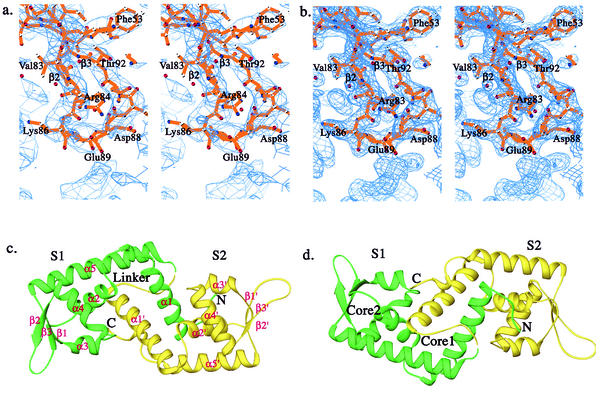
FIG. 1.
Overall structure of SarS. (a) The initial MIR map at the beta-hairpin region of S1 with the final SarS model, which is one of the most flexible parts in the entire SarS structure. (b) The final 2Fo-Fc map with the final SarS model at the same region as in panel a. (c) Ribbon diagram of the three-dimensional structure of the SarS protein. The first domain, S1, is shown in green. The second domain, S2, is yellow. (d) Orientation of the panel, 180°. Two hydrophobic cores are labeled. All figures were prepared using RIBBONS (6), except for Fig. 1a and b, which were prepared using BobScript (15).

