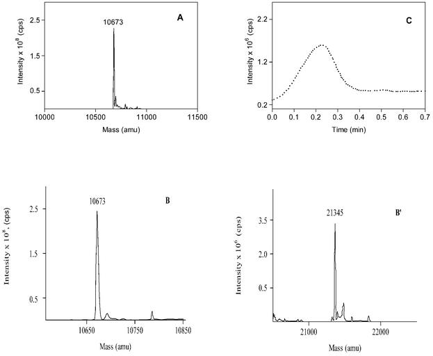
FIG. 5.
Mass spectra of M. tuberculosis Cpn10 in water-MeOH (1:1, vol/vol) at 4 μM (A) and 40 μM (B and B′), showing the existence of monomeric (B) and dimeric (B′) forms of the Cpn10 under these conditions. Panels B′ and B′′ are derived from the same spectrum and are separated for clarity due to the different intensities generated by the two molecular species. The solutions were infused into the spectrometer chamber as described in the experimental section. (C) Collision-induced dissociation of dimers to monomers obtained by increasing the mass spectrometer orifice voltage ramping from 5 to 120 V.

