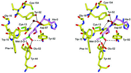
FIG. 8.
Stereoview of a putative model of Met-S-O binding to the active site prior to nucleophilic attack by Cys-13. Met-S-O-1, shown in violet, is modeled in the position of Met-1 of the crystal structure. Hydrogen bonds at a distance of 3.2 Å or less are shown as black dashed lines. This figure was generated using BOBSCRIPT (14), GLR (version 0.5.0; National Institutes of Health; http://convent.nci.nih.gov/∼web/glr/glrhome.html), and POV-Ray (version 3.5; POV-Team; http://www.povray.org).

