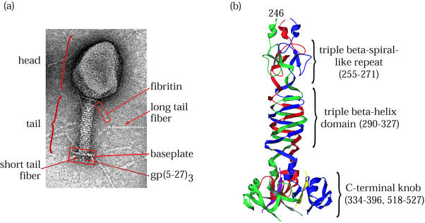
FIG. 1.
T4 virion and its short tail fiber, gp12. (a) Electron micrograph of bacteriophage T4 showing the locations of structural proteins and features. (b) Ribbon diagram of the T4 short tail fiber structure (67). The C-terminal domain at the bottom of the figure binds irreversibly to the bacterial host cell LPS. Residues 290 to 327 comprise a triple β-helix. A single β-strand motif similar but not identical to a triple β-spiral repeat is seen near the N terminus of the structure at the top. The three subunits are shown in red, green, and blue. (C-terminal strands whose connectivities were not assigned are shown in yellow, purple, and gray.)

