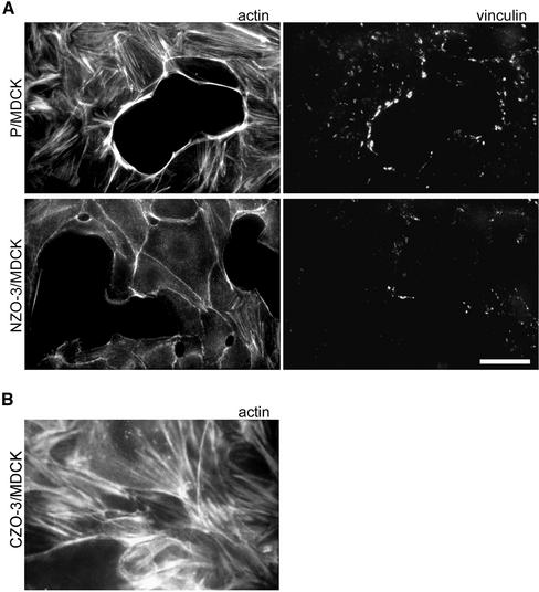
Figure 1.
NZO-3/MDCK cells have fewer stress fibers and focal adhesions than untransfected parental MDCK cells. (A) Parental MDCK (P/MDCK) and NZO-3/MDCK cells were grown to subconfluence and then costained with FITC-phalloidin to visualize F-actin, and anti-vinculin antibodies (rhodamine) as a marker for focal adhesions. NZO-3–expressing cells show a striking decrease in the number of stress fibers and a decrease in number and size of focal adhesions. Bar, 15 μm. (B) CZO-3/MDCK cells stained with FITC-phalloidin to visualize F-actin. These cells show the presence of stress fibers similar to parental MDCK cells.

