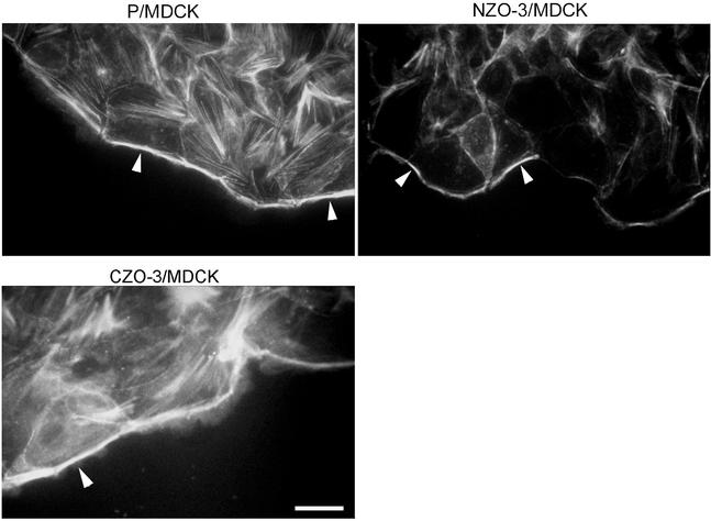
Figure 3.
NZO-3/MDCK cells have reduced F-actin staining at the free edge of the wound. Wounded monolayers were stained with rhodamine-phalloidin to visualize F-actin. Parental MDCK cells and CZO-3/MDCK cells show intense F-actin staining at free edge of cells adjacent to the wound (arrowheads). The amount of F-actin at the wound edge is less in MDCK/NZO-3 cells (arrowheads). Bar, 15 μm.

