Figure 1.
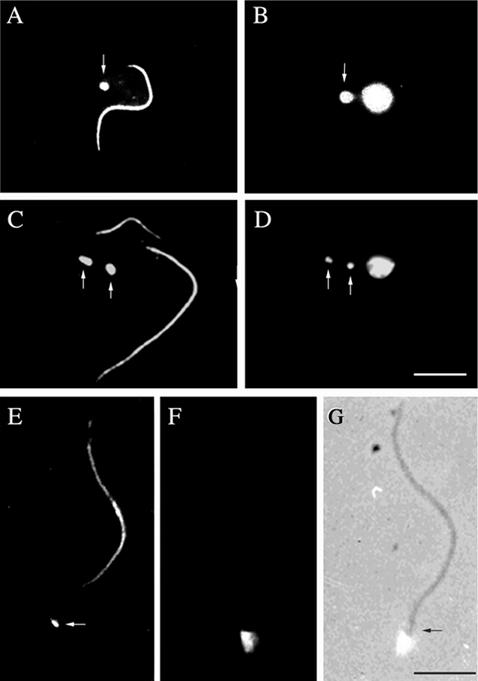
Kinetoplasts are physically attached to the flagellum basal bodies and are segregated by them during cell growth. Immunofluorescence of procyclic cells (A–D) or isolated flagella (E–G) by using two monoclonal antibodies, ROD 1 and BBA4, which illustrate the location of the paraflagellar rod within the flagellum in addition to the basal bodies, (arrows in A, C, and E). DAPI staining of the corresponding cells or flagella shows the location of nuclear and/or kinetoplast DNA (arrows in B and D). In E–G, the kinetoplast remains in contact with the isolated flagellum even after cell lysis and cytoskeleton depolymerization, illustrating the physical link between the kinetoplast and flagellum basal bodies via the TAC (E, F, and G). Bars, 5 μm.

