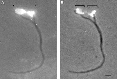
Figure 4.
Kinetoplast DNA replication and segregation occurs simultaneously in T. brucei procyclic cells. Procyclic cells were treated with BrdU to allow the replicating kinetoplasts to incorporate this proliferation marker. Cells were detergent extracted, the subpellicular microtubules depolymerized, and the resulting kDNA/flagella preparations probed with anti-BrdU antibodies. (A) kDNA/flagella complex visualized simultaneously with phase contrast and UV light by using the DAPI filter. The spread kDNA is seen lying between the basal bodies of a mature and immature flagellum (area below large bracket). The DAPI staining also shows the kinetoplast network to be in mid-to-late stages of replication as observed by the characteristic bilobed appearance. Anti-BrdU labeling combined with phase contrast of the same complex is shown in B illustrates that the network has two antipodal lobes of BrdU incorporation, located 180o apart (area below small brackets). Attachment of the flagella to the replicating network is clearly observed at the outermost poles of the network, within the sites where replicated DNA occurs. This is the site of the TAC. Bar, 1 μm.

