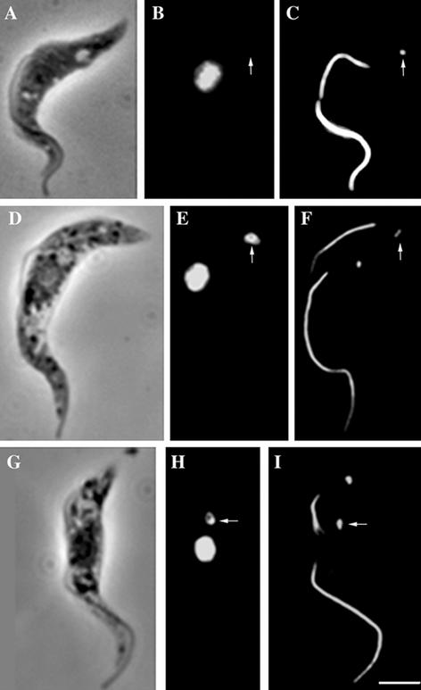
Figure 5.
Acriflavine blocks the postreplication mitochondrial genome linkage to the TAC. Phase contrast (A, D, and G), DAPI staining (B, E, and H), and immunofluorescence labeling (C, F, and I) of acriflavine-treated T. brucei cells was done using two monoclonal antibodies ROD 1 and BBA4. This indicates the location of the paraflagellar rod and the basal bodies after acriflavine treatment. DAPI staining of cells show the location of the nucleus and/or kinetoplast DNA. The cell in A, B, and C is dyskinetoplastic with no kDNA present (B, arrow). The ROD 1/BBA4 staining of this dyskinetoplastic cell shows the presence of a flagellum and a corresponding basal body (C, arrow). The cell in D, E, and F shows normal morphology when viewed by phase contrast microscopy, but the DAPI and immunofluorescence images show that the cell has one kinetoplast and two flagella. The single kinetoplast of this cell is associated with the new flagellum basal body (E and F, arrows). The cell in G, H, and I also has a “normal” morphology by phase contrast but again the DAPI and immunofluorescence images show that the cell has one kinetoplast and two flagella. In this case, the kinetoplast is associated with the old flagellum basal body. Bar, 5 μm.

