Figure 2.
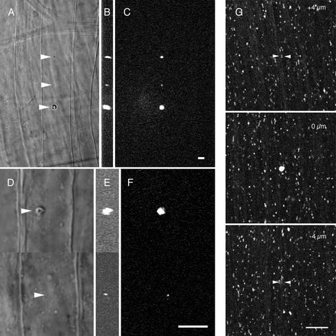
Small localized wounds in the squid fin nerve and giant axon. Different size wounds were made in a fin nerve of the squid with either a 25× 0.8 NA multi-immersion lens (A–C) or a 63× 1.2 NA (D–F) water immersion lens. Arrowheads point to the wound in the transmitted light images A and D. The corresponding fluorescent scar is shown in the x-y (C and F) and y-z (B and E) planes. The corresponding volumes for these fluorescent scars are 76, 10.9, and 376.1 μm3 (B and C) and 10.9 and 0.253 μm3 (E and F). (G) Spot wound in the squid giant axon using a 63× 1.2 NA water immersion lens. To confirm that damage was confined to the wound plane axonal transport was monitored by taking time series of Rhodamine 123 labeled mitochondria at the wound plane, 4 μm above and 4 μm below the wound plane (see online movies: above.mov and below.mov). The volume of the wound was 13 μm3. Arrowheads indicate the wound site in the out of plane images. Scale bar for all images, 10 μm.

