
Figure 4.
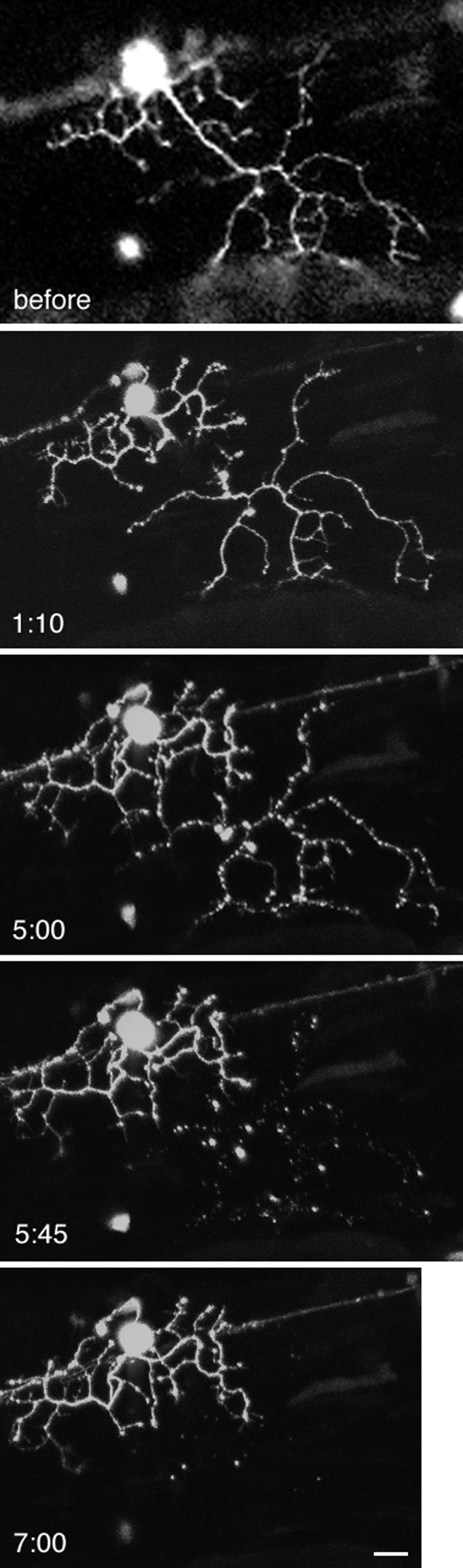
Severing of a neuronal dendrite. Injection of a transfection vector into fertilized zebrafish eggs resulted in mosaic labeling of neurons by GFP in 1.5-d-old embryos. A Rohon-Beard cell dendrite was severed by a single multiphoton line scan. Z series images were taken at 30–45-min intervals by confocal microscopy, with the time after wounding indicated on the figure. The severed dendrite remained intact for several hours. At 5 h, the beginning of degeneration was evident by the beaded appearance of the dendrite; the apparent connection at another point has not occurred and instead is due to the superposition of z series images. By 7 h, the remnants of the dendrite had disappeared. Scale bar, 10 μm.