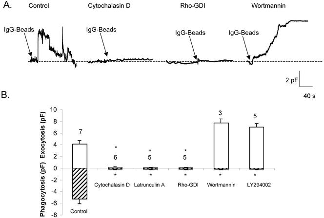
Figure 4.
Focal exocytosis is not affected by inhibitors of PI3 kinase but is blocked by actin polymerization antagonists. (A) Continuous Cm recordings from J774 macrophages incubated under control conditions or in the presence of cytochalasin D (0.5 μg/ml), latrunculin A (50 nM), Rho-GDI (10 μg/ml), or wortmannin (100 nM). All agents were preincubated with cells for 30 min before recording. In the case of Rho-GDI, 5–10 min was allowed to elapse to permit intracellular dialysis of the protein via the patch pipette before bead challenge. (B) Summary of mean peak Cm values (± SEM) obtained from cells incubated with actin polymerization antagonists or inhibitors of PI3 kinase (50 μM LY294002). Focal exocytosis (above baseline) and phagocytosis (below line) are shown; number of cells per condition indicated above bars. (Apparent “enhancement” of exocytosis by wortmannin and LY294002 is likely due to absence of contaminating phagocytosis, rather than to any genuine stimulatory effect.)

