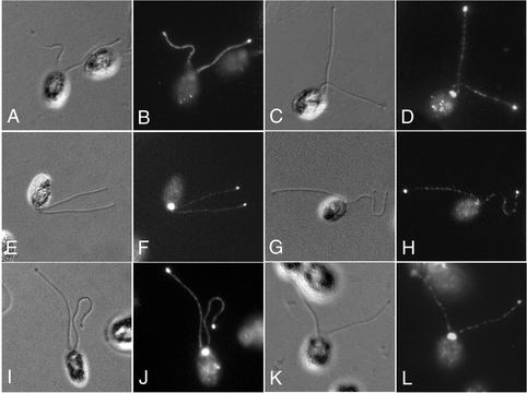
Figure 6.
Redistribution of IFT motors and IFT particles in a length control mutant. lf1 cells assemble flagella that are 2 to 3 times wild-type length (Barsel et al., 1988; Aselson et al., 1998). Small bulges are often visible at the tips of the lf1 flagella by differential interference contrast microscopy. lf1 cells were stained with antibodies to cDHC1b (A and B), LIC (C and D), p172, (E and F), p139 (G and H), and the FLA10 kinesin (I and J). Note the accumulation of all components at the flagellar tips (cDHC1b staining of the peribasal body region is out of the plane of focus in B). (K and L) Staining of lf3 cells with antibodies to the LIC.

