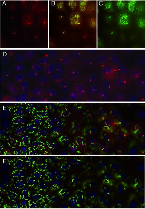
Figure 7.
D2LIC colocalizes with primary cilia in MDCK cells. Confluent cultures of polarized MDCK cells were stained with antibodies to D2LIC (shown in red) and either tubulin or the Golgi marker p115 (shown in green). The cells shown in A–C were costained with anti-D2LIC and anti-tubulin and then viewed from above, at the level of the apical cytoplasm and primary cilia. D2LIC staining (A), merged image (B), and tubulin staining (C). The cells shown in D–F were costained with anti-D2LIC, anti-p115, and Hoescht. D2LIC staining (D), merged image (E), and p115 staining (F). The Golgi apparatus is often out of focus in optimal views of the primary cilia. D2LIC is abundant in the apical cytoplasm, but clearly enriched in the primary cilia.

