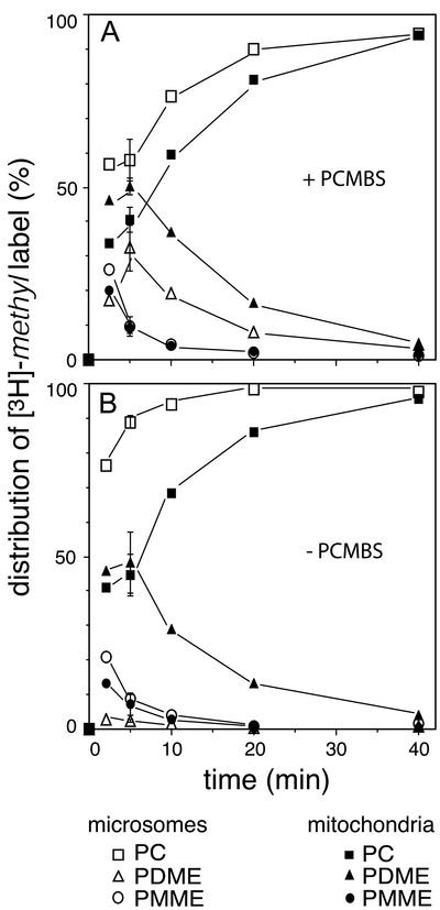
Figure 2.
The rates of accumulation of the [3H]-methyl label in PC in mitochondria and microsomes, as observed after subcellular fractionation in the presence (A) and absence (B) of 1 mM PCMBS in the breakage buffer. The distribution of the [3H]-methyl label over PMME, PDME, and PC, is presented as a percentage of the total amount of label incorporated in the phospholipids of microsomes and mitochondria. The error bars at t = 5 min reflect the SD (n = 3). For experimental conditions see MATERIALS AND METHODS.

