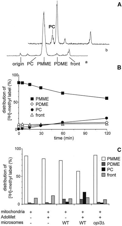
Figure 3.
Mitochondrial [3H]-methyl-PMME is converted to [3H]-methyl-PC by microsomal PLMT in vitro in the presence of AdoMet. (A) Radioactivity scan of a TLC plate showing the distribution of the [3H]-methyl label in a lipid extract of opi3Δ mitochondria before (a) and after (b) incubation for 2 h with wild-type microsomes and AdoMet. (B) Time course of the conversion of [3H]-methyl-PMME into [3H]-methyl-PDME and [3H]-methyl-PC. The distribution of the incorporated [3H]-methyl label over PMME, PDME, and PC, and the front is presented as percentage of the total amount of label incorporated into lipids. Data are averaged from four independent experiments with the SD not exceeding 4%. (C) Distribution of the [3H]-methyl label over PMME, PDME, and PC, and the front after 2 h of incubation of [3H]-methyl-PMME-loaded opi3Δ mitochondria in the absence and presence of microsomes and AdoMet as indicated.

