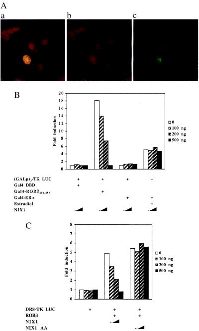
Figure 7.
(A) NIX1 is located in the nucleus. NIH 3T3 fibroblasts and HT22 hippocampal cells were transfected with pCMX-NIX1. NIX1 immunodetection with affinity-purified anti-NIX1 antibody and FITC-labeled secondary antibody is shown in green. DNA staining by using propidium iodide and RNase is shown in red. The superimposed fluorescence microscopy images of both results in a yellow, nuclear stain. (B and C) NIX1 suppresses transactivation of distinct agonist-bound nuclear receptors. (B) (GALp)3-TK LUC was transfected into NIH 3T3 cells together with increasing amounts of pCMX-NIX1 (100, 200, and 500 ng) and expression plasmids (100 ng each) encoding for Gal4 DBD alone or the indicated Gal4 DBD-receptor fusions. For Gal4-ERα, transfected cells were treated with 0.1 μM estradiol or vehicle. (C) HT22 cells were transfected with expression plasmids (100 ng) for RORβ and increasing amounts pCMX-NIX1 or pCMX-NIX1 AA (100, 200, 500 ng). DR8-TK LUC (500 ng) was used as reporter plasmids.

