Figure 1.
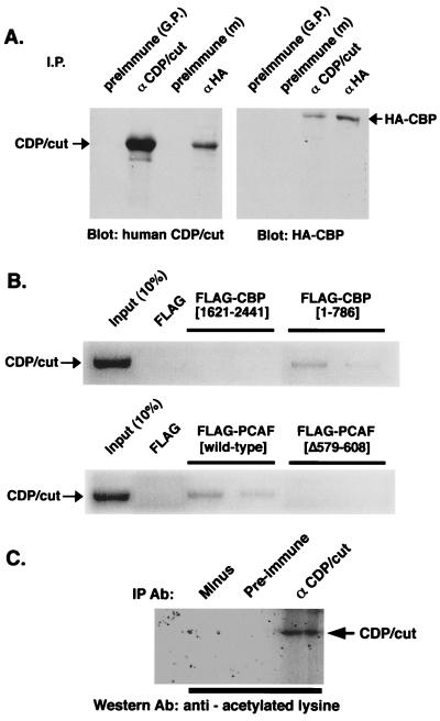
Interaction of CBP and PCAF with CDP/cut. (A) Immunoprecipitation/Western blot analysis of transfected HA-tagged CBP and CDP/cut. HeLa cells were cotransfected with expression vectors for CBP and CDP/cut. Immunoprecipitations with antibody or antiserum directed to either the HA-tag (αHA) or CDP/cut, respectively, from the lysates of transfected cells were recovered and subjected to immunoblotting by antisera indicated under the blot. Arrows indicate the predicted size of the detectable immunoblotted protein. (B) [35S]-labeled CDP/cut was synthesized in vitro and mixed with purified FLAG-tagged CBP, corresponding to the residues of CBP as indicated, immunoprecipitated with anti-FLAG M2 antibody (Sigma), electrophoresed, and detected by autoradiography. (Lower) [35S]-labeled CDP/cut was synthesized in vitro and mixed with purified FLAG-tagged PCAF (wild type) or PCAF (Δ579–608) and visualized as in Upper. (C) Acetylation of CDP/cut in vivo. Lysates from HeLa cells, transfected with pMT2-CDP, were immunoprecipitated with anti-CDP/cut antiserum or preimmune serum as a control. Immunoprecipitates were analyzed by immunoblot analysis by using a polyclonal antiacetylated lysine antibody.

