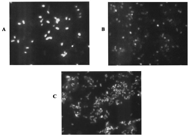
FIG. 3.
Microscopic analysis of fluorescence resulting from mixing of various test strains with 11.10/pMF3. The images in this figure have been magnified two times to clearly show differences in the response of the reporter. Cells in the mixture were (A) 11.10/pMF3 and Achromobacter cycloclastes, (B) 11.10/pMF3 and Rhizobium hedysari HCNT1, (C) 11.10/pMF3 and an environmental isolate. For A and C, the test strain (Achromobacter cycloclastes or an environmental isolate) was cultured microaerobically in medium supplemented with nitrate, harvested, resuspended, and transferred to a serum bottle. Cells of 11.10/pMF3 were added to each vial, and the vials were sealed. Each vial was supplemented with nitrate and incubated for a minimum of 2 h before samples were viewed under a microscope. For B, Rhizobium hedysari HCNT1 was cultured in medium lacking nitrogen oxides and mixed with the reporter strain as described above. In this case, nitrite rather than nitrate was added to the vial prior to incubation.

