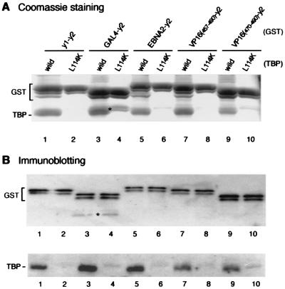
Figure 2.
Acidic activation domains interact with the concave surface of TBP. yTAND-1 (lanes 1 and 2) and various activation domains including GAL4 (lanes 3 and 4), EBNA2 (lanes 5 and 6), VP16 (residues 457–490 or 470–490; lanes 7–10) were fused to yTAND-2 (y2), and expressed as GST fusion proteins. These GST proteins were incubated with equimolar amounts of wild-type (odd lanes) or L114K mutant TBP (even lanes). After GST precipitation, the purified materials were analyzed by Coomassie blue staining (A) or immunoblotting with anti-GST and anti-TBP antibodies (B) as described in the Fig. 1 legend. Degraded GST proteins that closely migrated with TBP are indicated by asterisks.

