FIG. 4.
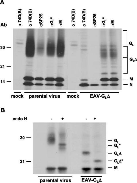
Immunoprecipitation analysis of EAV-GLΔ. EAV-GLΔ-infected, parental-virus-infected, and mock-infected BHK-21 C13 cells were labeled with [35S]methionine from 6.5 to 12 h p.i. in the absence (A) or presence (B) of DMJ. After removal of cell debris by low-speed centrifugation, the labeled virus present in the cell culture medium was dissociated in lysis buffer containing 20 mM NEM and subjected to immunoprecipitation. (A) Samples were immunoprecipitated either with the MAb 74D(B) or the antipeptide sera αGLC and αSP25 (all against GL) or with the antipeptide serum αM (directed against peptide SP06 derived from the M protein). (B) Samples were immunoprecipitated with αGLC. The immunoprecipitates were treated (+) or mock treated (−) with endo H as indicated. Samples were finally dissolved in Laemmli sample buffer containing 50 mM dithiothreitol and analyzed by SDS-15% polyacrylamide gel electrophoresis. Numbers on the left refer to positions of the molecular mass markers (in kilodaltons). Asterisks, positions of the deglycosylated GL polypeptides.

