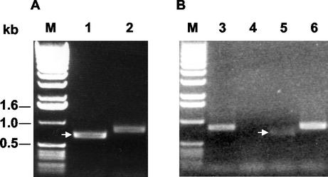
FIG. 5.
RT-PCR analysis of ORF5 from EAV-GLΔ inoculated into (A) and retrieved from (B) ponies. RT-PCR amplification of ORF5 was carried out on tissue culture fluid with primers JV1 and JV2. The presence of the deletion in ORF5 both for the input virus and the virus present in the blood of one of the ponies (7b69) was checked at day 4 after inoculation with EAV-GLΔ. Lane 1, EAV-GLΔ inoculum virus prepared in EEL cells; lanes 2 and 3, wild-type EAV strain CVL Bucyrus grown in RK13 cells; lane 4, uninfected RK13 tissue culture fluid; lane 5, virus recovered in RK13 cells from an EAV-GLΔ-infected pony (7b69); lane 6, challenge virus (LP3A+) prepared in EEL cells. On the left the positions and sizes of marker DNA fragments are indicated.

