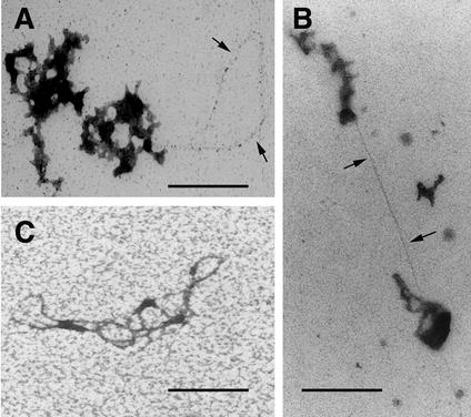
FIG. 6.
Visualization of nucleic acids in purified RTCs. Samples (3 and 4 h postinfection) were treated with proteinase K for 1 h (A) or 2 h (B), ethanol precipitated, adsorbed onto carbon-coated 400-mesh grids, washed in distilled water, and stained with 1% ethanolic UA diluted 1:5 in acetone for 5 min. (C) Bundle of nucleic acid filaments from a preparation deproteinized with guanidine thiocyanate. The arrows point to nucleic acids. Scale bar, 150 nm.

