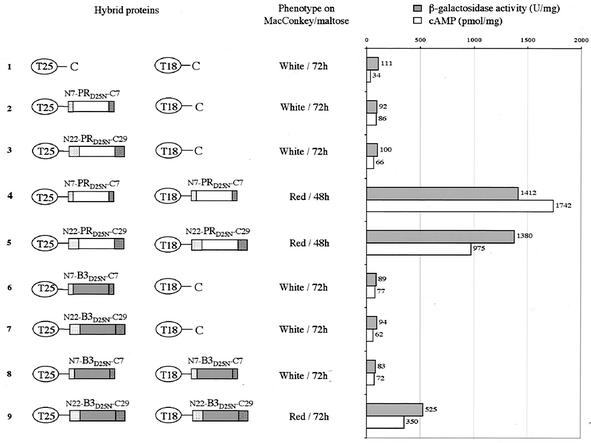
FIG. 5.
BACTH assay of HIV PR dimerization. The two AC fragments T25 and T18 are schematized by ovals, and the PR and B3 polypeptides are represented by white and grey rectangles, respectively. The N terminus HIV PR flanking sequences (TF derivatives: N7 and N22) are represented as dotted rectangles, whereas C terminus flanking sequences (RT derivatives: C7 and C29) are represented as hatched rectangles. The phenotype of DHT1 cells expressing the indicated pairs of proteins was scored on MacConkey-maltose plates plus kanamycin and ampicillin and supplemented with 0.5 mM isopropyl β-d-thiogalactoside (IPTG). β-Galactosidase activities and cAMP levels were measured on liquid cultures grown overnight at 30°C in LB supplemented with appropriate antibiotics and 0.5 mM IPTG. The plasmids expressing the indicated hybrid proteins were as follows: lane 1, pKT25 + pST18C; lane 2, pKT25HIV-S + pST18C; lane 3, pKT25PR-L + pST18C; lane 4, pKT25PR-S + pST18CPR-S; lane 5, pKT25PR-L + pST18CPR-L; lane 6, pKT25B3-S + pST18C; lane 7, pKT25B3-L + pST18C; lane 8, pKT25B3-S + pST18CB3-S; lane 9, pKT25B3-L + pST18CB3-L.

