FIG. 4.
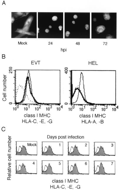
Expression of class I MHC molecules in HCMV-infected EVT. (A) Indirect immunofluorescence analysis. Expression of class I MHC molecules in HCMV-infected EVT. EVT were infected with HCMV at an MOI of 5 and examined at 72 hpi using the MAb W6/32, which reacts with HLA-A, -B, -C, -E, and -G. (B) Flow cytometric analysis of the surface expression of class I MHC molecules at 72 hpi. EVT express HLA-C, -E, and -G. HEL express HLA-A and -B. These class I MHC molecules of HCMV-infected cells (thick line) and mock-infected cells (thin line) were detected using the MAb W6/32 or isotype control (dotted line). (C) Expression of class I MHC molecules in HCMV-infected EVT on later days. The flow cytometry of EVT was performed for 7 days after infection. Shaded curve, W6/32 antibody; open curve, isotype control antibody.

